liver enzymes, who have subsequently been shown to have liver abscesses. I have
been asked to help find the cause of the liver abscess and advise on treatment. Interestingly...the cause can usually be determined from the history taking alone - laboratory tests just provide confirmation. Although liver abscesses are
not particularly common, they are good examples of how a careful and systematic approach can elicit the cause and lead to the correct treatment.
commonly in my clinical work:
1. Polymicrobial bacterial infection due to bacteria ascending the biliary tract – most common in the UK
2. Parasitic infection due to parasite ascending the biliary tract – more common in returned travellers or people born overseas
In polymicrobial bacterial infections there is usually an
underlying problem with the biliary tract in the first place such as:
• Obstruction to bile drainage e.g. gallstones
• Abnormal anatomy which allows bacteria easier access e.g. tumour distorting the common bile duct
• Surgical alteration of the common bile duct such as following a sphincterotomy or insertion of a stent
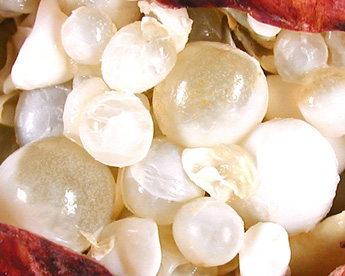
In parasitic infections there is usually a history of foreign travel, ingestion of contaminated food or water or occupational exposure. The two main parasites are Amoebae and Echinococcus (see image of multiple hydatid cysts in Echinococcosis).
A thorough clinical history (regarding gallstones, biliary tract
surgery, travel history, pets or occupational history) can identify the most likely cause. This should be combined with microbiological culture of blood and pus, as well as serum for amoebiasis and echinococcosis. Radiological detection
of a cause of obstruction (in polymicrobial bacterial infections) and the monitoring of how both (polymicrobial or parasitic) respond to therapy is important.
Treatment
It is essential to make the distinction between the causes
because the treatments are very different.
Polymicrobial bacterial liver abscesses require surgical drainage (if possible) followed by 2-4 weeks of
broad-spectrum antibiotics (such as combinations of Amoxicillin, Gentamicin and Metronidazole or Co-amoxiclav PLUS Gentamicin) until clinical and radiological resolution.
Amoebic abscesses should be drained if they are more than 5cm diameter and amenable to drainage, with PO Metronidazole 800mg TDS for 10 days followed by PO Diloxanide Furoate 500mg TDS for 10 days (adults).
Echinococcus is much more complicated and difficult to treat. The danger is that if the hydatid cysts rupture during surgery the patient often experiences an anaphylactic reaction and goes on to develop multiple secondary cysts within the peritoneum, with bowel obstruction due to adhesion formation.
The World Health Organisation recommends 4 possible options for treatment:
- PAIR - percutaneous treatment of the hydatid cysts with Puncture, Aspiration, Injection, Re-aspiration if multiple cysts (NB it is PAIR not pears!)
- Surgical removal if single cyst
- Drug treatment with Mebendazole, Albendazole or
Praziquantel - Observation if small cyst and asymptomatic
PAIR is a minimally invasive method but it requires considerable expertise. Each cyst is drained with a fine needle or catheter, followed by the injection of a drug such as hypertonic saline or ethanol to kill the protoscolices (immature forms of the Echinococcus) with final drainage of the hypertonic saline or ethanol. PAIR is usually now combined with PO Albendazole therapy at 400mg BD for 1 week before PAIR and 4 weeks after PAIR to prevent
recurrence or secondary cysts.
Although doctors in the UK are more likely to see polymicrobial bacterial more frequently (3-4 per 100,000 population) than parasitic abscesses from either amoebae
(1-2 per 100,000 population) or a canine related Echinococcus (<1 per 100,000 population) all should be part of the differential diagnosis of patients with liver abscesses. Just think if you identified an Echinococcus liver abscess from the history it would certainly impress the microbiologist as well as benefit the patient.

 RSS Feed
