“I have a patient with a bright red nose” said the junior Doctor.
The Microbiologist double-checked his calendar to make sure it wasn’t March 15th and the junior performing a joke for Comic Relief’s “Red Nose Day”, a charity day here in the UK!
“What do you mean a bright red nose” the Microbiologist asked, adding “it’s not Comic Relief Day, you know?” heading off the “fooled you” prank.
“What!?”... The junior inhaled deeply, ignored the Microbiologist, and continued “It’s a kind of reddish purple and very painful. It looks a bit like cellulitis but it is in a very odd place, and the patient doesn’t have a fever. What antibiotics do you think we should give?”
Muttering, “this isn’t just a dial an antibiotic line”, the Microbiologist decided he would like to see this patient’s nose and find out what was going on himself before advising what treatment should be started…
He added “your patient needs a diagnosis before throwing random drugs at them to see if they get better”.
As the Microbiologist walked into the patient’s room to introduce himself he couldn’t help but see that the patient did indeed have a bright red nose. He chuckled to himself; the patient definitely didn’t need one of those plastic red noses.
After a brief embarrassed pause the Microbiologist finally introduced himself. The patient smiled and then unconsciously reached up and stroked his nose with his thumb and forefinger.
With sudden understanding the Microbiologist asked “do you have anything to do with animals, either work or at home?”
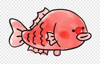
“I am a keen keeper of Koi carp and have just finished transferring them to their new pond… I designed it myself” the patient said proudly.
It was the Microbiologists turn to smile. “You have erysipeloid” he told the patient confidently.
“Eh, sip, what? No, I have Koi carp, they’re fish!” said the patient, confused.
The Microbiologist double-checked his calendar to make sure it wasn’t March 15th and the junior performing a joke for Comic Relief’s “Red Nose Day”, a charity day here in the UK!
“What do you mean a bright red nose” the Microbiologist asked, adding “it’s not Comic Relief Day, you know?” heading off the “fooled you” prank.
“What!?”... The junior inhaled deeply, ignored the Microbiologist, and continued “It’s a kind of reddish purple and very painful. It looks a bit like cellulitis but it is in a very odd place, and the patient doesn’t have a fever. What antibiotics do you think we should give?”
Muttering, “this isn’t just a dial an antibiotic line”, the Microbiologist decided he would like to see this patient’s nose and find out what was going on himself before advising what treatment should be started…
He added “your patient needs a diagnosis before throwing random drugs at them to see if they get better”.
As the Microbiologist walked into the patient’s room to introduce himself he couldn’t help but see that the patient did indeed have a bright red nose. He chuckled to himself; the patient definitely didn’t need one of those plastic red noses.
After a brief embarrassed pause the Microbiologist finally introduced himself. The patient smiled and then unconsciously reached up and stroked his nose with his thumb and forefinger.
With sudden understanding the Microbiologist asked “do you have anything to do with animals, either work or at home?”
“I am a keen keeper of Koi carp and have just finished transferring them to their new pond… I designed it myself” the patient said proudly.
It was the Microbiologists turn to smile. “You have erysipeloid” he told the patient confidently.
“Eh, sip, what? No, I have Koi carp, they’re fish!” said the patient, confused.
What is erysipeloid?
Erysipeloid is a form of cellulitis caused by the bacterium Erysipelothrix rhusiopathiae. Patients have a single extremely painful violaceous (violet coloured) lesion, usually on a finger, with associated inflammation and enlargement of the local lymphatic system (lymphangitis). However about 10% of these patients also have a low grade fever and pain in the joints (arthralgia). More rarely erysipeloid can spread to cause a diffuse rash which is always associated with a fever and arthralgia; this presentation is often prolonged and relapsing in nature.
In immunocompromised patients, although occasionally in immunocompetent patients as well, erisypeloid can become a systemic infection with E. rhusiopathiae spreading to other body sites such as heart valves causing endocarditis, bone, joints, muscle and brain.
It is important not to confuse the term erysipelas, which is a type of skin infection caused by the Group A beta-haemolytic streptococcus (Streptococcus pyogenes) in which fever and raised inflammatory markers are much more common. They do look very similar though and fortunately (if you do not bother to distinguish… I mean diagnose between them!) both tend to respond to the same antibiotics. It’s important to differentiate because Group A beta-haemolytic streptococcus or erysipelas is associated with post-streptococcal sequelae such as rheumatic fever, rheumatic heart disease and glomerulonephritis whereas erysipeloid is not.
Where does Erysipelothrix rhusiopathiae come from?
Given the flavour of recent blogs it may come as no surprise to know that E. rhusiopathiae is a zoonotic organism. It is an organism that normally infects pigs, causing “swine fever”, but it can also be found in the slime covering fish (in whom it doesn’t cause infection), in poultry and in sheep. It is major problem for the swine meat industry as it can cause major outbreaks and severe disease in herds of pigs.
Infection in humans with E. rhusiopathiae is most commonly seen in fishermen, those who keep fish, farmers, abattoir workers, butchers and veterinary surgeons.
Just so you know, and so I can justify my collection of antique medical textbooks, the Father of Microbiology, Robert Koch, was the first to isolate E. rhusiopathiae back in 1878. Louis Pasteur then managed to grow it from pigs in 1882, and Friedrich Loeffler went on to show it caused swine fever in 1886. It wasn’t until 1909 that they realised it also caused human disease.
How is erysipeloid diagnosed?
The diagnosis of erysipeloid is usually clinical, based on the appearance of the skin lesion in someone with appropriate animal exposure. If confirmation is required biopsy specimens should be taken from the edge of the skin lesion (NB superficial swabs are not suitable as the organism is often deep in the tissue); in febrile patients also take blood cultures.
E. rhusiopathiae is a Gram-positive, catalase negative bacillus that produces hydrogen sulphide. These two aspects help distinguish it from other important Gram-positive bacilli such as Listeria monocytogenes (which is catalase positive and doesn’t produce hydrogen sulphide). Although clinically similar, Group A beta-haemolytic streptococcus (Streptococcus pyogenes) is actually a Gram-positive coccus and so looks different to E. rhusiopathiae when looked at down a microscope.
It isn’t difficult to grow E. rhusiopathiae in the lab. It grows easily on blood agar at 37oC as a facultative anaerobe (grows both aerobically and anaerobically). Colonies tend to be small and slow growing and pretty non-descript; they are often alpha-haemolytic (S. pyogenes is beta-haemolytic). The main issue with growing E. rhusiopathiae is if there are faster growing bacteria mixed in, as these will grow over the top and you won’t notice the small Gram-positive hiding underneath.
How is erysipeloid treated?
Erysipeloid is usually a self-limiting problem getting better within a few weeks, although antibiotics speed up resolution and are usually given to relieve the pain.
The normal first line treatments of infections with E. rhusiopathoiae are beta-lactams such as penicillins or cephalosporins. Other antibiotics that are active include carbapenems, Clindamycin, Linezolid, Ciprofloxacin and Daptomycin.
Treatment duration should be based on the type of clinical picture e.g. 10-14 days for skin infections, 4 weeks for endocarditis and 6 weeks for osteomyelitis.
NOTE: Vancomycin and Teicoplanin are usually active against Gram-positive bacteria and are often used to treat infections caused by these Gram-positive bacteria however E. rhusiopathiae is an exception to this rule as it is resistant to these antibiotics. It is also resistant to aminoglycosides such as Gentamicin which is often used to treat sepsis and so if you rely on using these antibiotics to treat patients with systemic infections caused by E. rhusiopathiae they won’t work.
Nowadays there is an animal vaccine for use in pigs to prevent swine fever, but no vaccine for humans. Anyone handling potentially infected or colonised animals should wear gloves and observe good hand hygiene.
So the patient was started on penicillin and his painful swollen nose quickly settled down. He also bought a nice new pair of waterproof gloves to help with future movement of his fish.
The Microbiologist wandered off the ward wondering what was for lunch this Friday… he expected it was his favourite, fish and chips.
 RSS Feed
