A sheep farmer presented to the hospital with a persistent cough and fever. A chest x-ray showed minor patchy consolidation but as the patient was otherwise fit and well he was quite rightly allowed to go home with a course of PO Amoxicillin (1st line treatment for community acquired pneumonia). A week later the patient was still feeling unwell so he went to his GP who changed him to PO Co-amoxiclav…just in case the bacteria were resistant. Five days later the patient returned to the hospital and was changed again to IV Piptazobactam as he was still no better and the doctors thought the oral antibiotics weren’t working.

Honest... these are real! Search "M8 red sheep"
Following the post-admission ward round the junior doctor rang the Duty Microbiologist with the question “I have a query about a patient with a fever and pneumonia, could I get some advice please?” Having heard the patients’ history the Microbiologist answered “what you’re asking is whether the patient has a ‘Query’ fever?”… “Yes, yes, I’ve already told you the patient has a fever, a temperature of 39oC” said the junior a little puzzled by the odd response. “Query fever” said the Microbiologist again pausing then adding “your diagnosis…its sounds like Query fever, you might recognise it by its abbreviated name…simply Q fever”. The junior paused, he had heard of Q fever but never knew the “Q” stood for “Query”! The Microbiologist broke the silence and said “take some blood samples to look for antibodies and change to PO Doxycycline… oh and thanks I’ll use Q fever for this week’s blog” The junior was left thinking “Microbiologists... surely they make this stuff up!”
The results of the blood tests confirmed that the patient did in fact have Q fever. So what is Q fever? That’s a very interesting query… okay, sorry, I’ve been told time and again not to use the word “interesting” to describe anything microbiological but I couldn’t resist!
Q fever is the name given to infection caused by the bacterium Coxiella burnetii; the “Q” really does stand for “Query” as the cause of the fever in the original 1935 outbreak in abattoir workers in Australia was unknown. Q fever occurs all over the world with a few exceptions, apparently there is no Q fever in New Zealand, possibly because New Zealand has no native mammals and therefore did not originally have any animals to act as hosts for the ticks which are the main reservoir of C. burnetii.
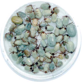
Q fever is a zoonotic infection; it comes from contact with animals, especially farm animals such as cattle, goats and sheep (although most mammals including household pets can be infected). Infected mammals excrete the bacterium in body fluids such as urine, faeces and milk as well as it being present in infected animal tissues. Those most at risk of infection are those who might have contact with infected animals or their body fluids:
Acute infection
The clinical presentation of Q fever following an incubation period of about 20 days is very variable. Up to 50% of patients are asymptomatic. The most common acute presentations of Q fever are:
Symptomatic infection is more likely to occur in adults, with adults more likely to have pneumonia or endocarditis and children hepatitis. Oddly, the clinical presentation also tends to vary on where in the world infection was acquired; in Canada pneumonia is more common whereas in France and Spain hepatitis is the usual presentation.
Chronic infection
Chronic infections occur in up to 5% of patients following both symptomatic and asymptomatic acute infection with Q fever. Chronic infections are more likely to occur in those with underlying immunosuppression, pregnancy, pre-existing cardiovascular disease or prosthetic joints. The bacterium lives and continues to replicate within the hosts macrophages (which renders them unable to kill the bacterium, as it would be self-destructive) resulting in a sustained bacteraemia. There are also high levels of autoantibodies and these cause further damage to tissues.
The most common chronic infections are:
How do you diagnose Q fever?
C. burnetii is a bit of a nuisance when it comes to diagnosis as it doesn’t grow in routine blood cultures or on agar; it can only survive inside other cells (a bit like a virus). In order to make a diagnosis you have to look for the body’s immune response to infection by detecting antibodies against C. burnetii.
There are 2 main types of antibody against C. burnetii, phase I and phase II. Phase I antibodies are produced when the bacterium is highly infectious and capable of infecting humans whereas phase II is produced when the bacterium has undergone antigenic shift into a non-infectious form (it has changed the antigens it is expressing rather than changing its genetics as with Influenza). The different phases of antibody can be used to differentiate acute and chronic infection but be warned the results are counter intuitive. Phase I DOES NOT go with acute infection and phase II with chronic infection as you would imagine! It feels wrong but it is the way it is!!
- Animal farmers and abattoir workers
- People who live downwind of animal farms where contaminated manure, straw or dust blow across their houses and gardens
- People who live in endemic countries e.g. French Guiana
Acute infection
The clinical presentation of Q fever following an incubation period of about 20 days is very variable. Up to 50% of patients are asymptomatic. The most common acute presentations of Q fever are:
- Self-limited flu-like illness – high fever (up to 40oC), tiredness, headache and muscle pains are common and typically last for 1-3 weeks
- Pneumonia – usually mild with a non-productive cough and fever lasting anywhere from 10 days to 3 months, mortality is approximately 1%
- Hepatitis – raised liver enzymes, hepatomegaly and fever
- Endocarditis – this is thought to be an autoimmune complication of acute infection in about 1% of patients and occurs in the presence of antiphospholipid antibodies which can be detected in the patients’ blood
- Rash – occurs in 10% of patients and can be maculopapular or purpuric
- Pericarditis and myocarditis – this rare complication occurs in less than 1% of patients with acute Q fever but has a high mortality (25%)
- Meningitis or encephalitis – occurs in less than 1% of patients
Symptomatic infection is more likely to occur in adults, with adults more likely to have pneumonia or endocarditis and children hepatitis. Oddly, the clinical presentation also tends to vary on where in the world infection was acquired; in Canada pneumonia is more common whereas in France and Spain hepatitis is the usual presentation.
Chronic infection
Chronic infections occur in up to 5% of patients following both symptomatic and asymptomatic acute infection with Q fever. Chronic infections are more likely to occur in those with underlying immunosuppression, pregnancy, pre-existing cardiovascular disease or prosthetic joints. The bacterium lives and continues to replicate within the hosts macrophages (which renders them unable to kill the bacterium, as it would be self-destructive) resulting in a sustained bacteraemia. There are also high levels of autoantibodies and these cause further damage to tissues.
The most common chronic infections are:
- Endocarditis – usually associated with low-grade fever, tiredness, weight loss, and night sweats. It occurs between 2 months and 2 years after the original infection and is more common in the presence of a pre-existing heart valve abnormality
- Vascular infection – presents with fever, abdominal pain and weight loss. There is usually a pre-existing aneurysm or vascular graft
- Bone and joint infection – presents with fever and joint pain in the same way as any other joint infection and can occur in both native and prosthetic joints
How do you diagnose Q fever?
C. burnetii is a bit of a nuisance when it comes to diagnosis as it doesn’t grow in routine blood cultures or on agar; it can only survive inside other cells (a bit like a virus). In order to make a diagnosis you have to look for the body’s immune response to infection by detecting antibodies against C. burnetii.
There are 2 main types of antibody against C. burnetii, phase I and phase II. Phase I antibodies are produced when the bacterium is highly infectious and capable of infecting humans whereas phase II is produced when the bacterium has undergone antigenic shift into a non-infectious form (it has changed the antigens it is expressing rather than changing its genetics as with Influenza). The different phases of antibody can be used to differentiate acute and chronic infection but be warned the results are counter intuitive. Phase I DOES NOT go with acute infection and phase II with chronic infection as you would imagine! It feels wrong but it is the way it is!!

Click for larger image
Q fever can also be diagnosed by using PCR to detect C. burnetii in tissue (e.g. heart valve, bone, joint fluid, etc.) and can be helpful in the small number of patients who are strongly suspected to have Q fever but in whom the antibody tests are negative. PCR is not routinely performed because it requires an invasive procedure to get tissue and can still be prone to false negative results (up to 30% in some studies) especially when taken after 10 days in acute infection.
Okay, so you’ve made the diagnosis, how do you treat your patient?
The treatment of Q fever depends on whether it is acute or chronic and can take up to 2 years in order to completely eradicate the bacterium. In adults the choices are usually:
Okay, so you’ve made the diagnosis, how do you treat your patient?
The treatment of Q fever depends on whether it is acute or chronic and can take up to 2 years in order to completely eradicate the bacterium. In adults the choices are usually:

Click for larger image
Note: whilst Q fever is not a specifically named notifiable disease in the UK, Public Health England should be informed about cases as outbreaks can and have occurred in the past in relation to animal and farm exposure.
So the patient underwent echocardiography which fortunately showed no pre-existing heart disease. He continued on Doxycycline for 14 days and made a full recovery. Failure to diagnose and treat the initial acute infection can lead to the development of chronic Q fever. The public health team were able to confirm that there had been no other cases relating to the same farm but a general reminder was sent to all the local hospitals asking them to be vigilant for further cases.
So the patient underwent echocardiography which fortunately showed no pre-existing heart disease. He continued on Doxycycline for 14 days and made a full recovery. Failure to diagnose and treat the initial acute infection can lead to the development of chronic Q fever. The public health team were able to confirm that there had been no other cases relating to the same farm but a general reminder was sent to all the local hospitals asking them to be vigilant for further cases.
 RSS Feed
