“I have a patient with facial cellulitis but they’re not responding to antibiotics, my Consultant has asked me to find out if there is a stronger antibiotic we can give” said the oncall doctor.
The Microbiologist rolled his eyes; why do people persist in thinking about antibiotics in terms of “stronger” when what they really mean is “broader spectrum”!
“Why don’t you start by telling me something about the patient?” said the Microbiologist through gritted teeth.
“Oh, okay” said the Doctor, thinking this was just going to waste more of his precious time.
“She has just returned from trekking in Bolivia where they stayed in mud huts. The patient thinks they might have been bitten on the face as there were quite a lot of big beetles around. She now has a swollen right eye and we’ve been giving IV Teicoplanin but it hasn’t made any difference so we want to give her something… stronger.”
His curiosity piqued, the Microbiologist let the latest “stronger” slip past.
“Are both upper and lower eyelids swollen? What did the insects look like exactly?”
“Yes, both eyelids are swollen and I have no idea what the bugs looked like” said the Doctor, thinking this was taking entirely too long.
“Okay, this sounds like Romana’s sign. I think your patient might actually have Chagas disease. Have you done a blood film? You need to send us blood for PCR and in the meantime I’ll pop over and see if your patient can describe the bugs in more detail”.
“Huh, so there is no antibiotic choice then?” grunted the Doctor thinking he wasn’t going to get that stronger antibiotic after all… and got the impression the Microbiologist was going off on a tangent which he wasn’t going to know much about!
What is Chagas disease?
Chagas disease, also known as American trypanosomiasis, is caused by the parasite Trypanosoma cruzi. Most mammals can be infected by T. cruzi including humans, rodents, cats, dogs and cattle.
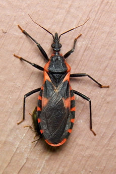
Infection is spread by an insect vector, usually the Triatomine bugs (also known as Kissing, Reduviid or Assassin bugs). While the bug is feeding it defecates and leaves it’s faeces to either penetrate the bite wound or, if on the face, it penetrates through the conjunctiva… yep it poos in your eye! Gross….
Chagas disease can also be spread via blood transfusion from an infected donor as well as congenital infection from an infected mother to her child (the risk of congenital infection from an infected mother is up to 10%). Many countries have systems in place to restrict blood donation from those who have either lived in or visited endemic countries. Less commonly infection can occur from transplanted organs or via ingestion of food or water contaminated with Reduviid bug faeces.
Apparently Chagas disease is responsible for the most illnesses and deaths of any parasite in the Western hemisphere, including more than malaria! Chagas disease occurs throughout the Americas from the Southern United States to the North of Argentina and Chile, with an estimated 6 million cases. The highest prevalence of Chagas disease is in Bolivia, Argentina, Paraguay, Ecuador, El Salvador, and Guatemala.
Traditionally Chagas disease was an infection of people in rural areas where the insect vector lived in cracks in the walls, or the thatched roofs, of adobe houses. However with increasing population movement both internally and globally Chagas disease acquired rurally can present at a later date in urban environments as well as in non-endemic countries. Control programs have eliminated the insect vector in countries like Brazil meaning that the proportion of congenital infections is now higher than naturally acquired infection.
How does Chagas disease present?
The incubation period for Chagas disease is usually 1-2 weeks but can be up to 4 months in transfusion transmission.
Most acute infections (within 8-12 weeks) with T. cruzi occur in children and are asymptomatic or very mild. Others experience mild fever, lymphadenopathy, malaise and hepatosplenomegaly (enlarged liver and spleen). A few patients get a swelling at the site of inoculation, known as a Chagoma; if this occurs after conjunctival inoculation causing swelling of the eye lids then it is known as Romana’s sign. Serious acute infection with myocarditis or meningoencephalitis occurs in less than 1% of patients.
However the most serious manifestations of Chagas disease, in 20-40% of patients, present up to 30 years later with either cardiac or less commonly gastrointestinal disease.
Cardiac Chagas disease usually presents with progressive heart failure, shortness of breath, fatigue, pulmonary and peripheral oedema (fluid accumulation) and chest pain. The heart becomes dilated and there is a risk of arrhythmias and emboli as a result of the heart not beating properly. The prognosis from cardiac disease is poor with over 30% dying from sudden cardiac death within 8 years from diagnosis. Others will die young from progressive heart failure; although there are scoring systems, e.g. the Rassi score, for trying to work out who will die and when, these are not that reliable.
Gastrointestinal Chagas disease is due to an autoimmune cross reaction triggered by antibody against the parasite which damages the nerves supplying the gut, especially the oesophagus and distal colon. Loss of innervation leads to progressive dilatation and eventually mega-oesophagus and mega-colon. Patients are unable to swallow safely and are at risk of aspiration and choking; they also become unable to absorb food properly, develop bowel obstruction and malnutrition.
How is Chagas disease diagnosed?
The easiest way to diagnose acute Chagas disease is to look at a film of blood and see the parasite in the red blood cells. The parasitaemia lasts about 90 days; in chronic infections the film will be negative as the parasites have been cleared from the bloodstream. Acute Chagas disease is now normally diagnosed using PCR on whole blood.
Chronic Chagas disease is difficult to diagnose and requires demonstration of IgG against T. cruzi. Because none of the tests are particularly good, and are prone to false positives, two different tests based on different antibody reactions should be used and the diagnosis made if BOTH are positive AND the patient has evidence of myocarditis or gastrointestinal disease.
Patients treated for Chagas disease should have follow up serology; the IgG should reduce with successful treatment. If the amount of IgG does not reduce then the patient may require retreating.
How is Chagas disease treated?
There are 2 parts to the treatment of Chagas disease; treatment of the parasitic infection and treatment of the chronic cardiac and gastrointestinal complications.
Antiparasitic drugs are used for both acute and chronic Chagas disease. There are two drugs; Benznidazole and Nifurtimox. Benznidazole tends to be first line treatment as it is better tolerated with fewer side effects. Treatment in acute infection reduces the duration and severity of symptoms (if present) and clears the parasitaemia resulting in cure in 60-85% (>90% for congenital infection if treated in the first year of life).
Treatment with antiparasitic drugs in the chronic form can reduce progression of cardiac and gastrointestinal disease by about 70-80%, especially if given before symptoms have progressed too far towards severe heart failure, mega-oesophagus or mega-colon.
Cardiac disease is treated in the same way as heart failure or arrhythmias for other causes with diuretics, beta-blockers and anticoagulants. Gastrointestinal disease is managed with dietary changes but ultimately patients may require extensive surgery with oesophagectomy and colectomy.
Prevention
The best way for travellers to prevent infection with T. cruzi is to avoid being exposed to the insect vector, sleeping under insecticide impregnated nets, not staying in rustic accommodation and avoiding street food that might be contaminated with insect faeces. Essentially travellers can avoid infection by normal simple common sense “safe traveller” behaviour.
For people living in endemic countries the same measures for prevention apply. Many South American countries have embarked on programs to improve rural housing by replacing adobe huts with brick and corrugated iron buildings… not as pretty maybe but considerably safer.
Congenital infections can be reduced by screening pregnant women by PCR as negative PCR correlates with very low or absent parasites in blood and therefore substantially reduced risk of in utero infection. There are no drugs that can be used safely in pregnancy to reduce the risk of transmission in PCR positive women. You have to wait for the child to be born before trying to treat the child and the mother.
So the Microbiologist spoke to the patient who described the Reduviid bugs in great detail (apparently they are 2cm long but quite pretty!). A film was looked at that day by the haematologists who saw parasites in the blood film. With the diagnosis confirmed the patient was started on Benznidazole and follow up blood tests fortunately showed a complete cure, although they had developed a phobia to big creepy bugs… but I guess that was to be expected really!
“Okay, this sounds like Romana’s sign. I think your patient might actually have Chagas disease. Have you done a blood film? You need to send us blood for PCR and in the meantime I’ll pop over and see if your patient can describe the bugs in more detail”.
“Huh, so there is no antibiotic choice then?” grunted the Doctor thinking he wasn’t going to get that stronger antibiotic after all… and got the impression the Microbiologist was going off on a tangent which he wasn’t going to know much about!
What is Chagas disease?
Chagas disease, also known as American trypanosomiasis, is caused by the parasite Trypanosoma cruzi. Most mammals can be infected by T. cruzi including humans, rodents, cats, dogs and cattle.
Infection is spread by an insect vector, usually the Triatomine bugs (also known as Kissing, Reduviid or Assassin bugs). While the bug is feeding it defecates and leaves it’s faeces to either penetrate the bite wound or, if on the face, it penetrates through the conjunctiva… yep it poos in your eye! Gross….
Chagas disease can also be spread via blood transfusion from an infected donor as well as congenital infection from an infected mother to her child (the risk of congenital infection from an infected mother is up to 10%). Many countries have systems in place to restrict blood donation from those who have either lived in or visited endemic countries. Less commonly infection can occur from transplanted organs or via ingestion of food or water contaminated with Reduviid bug faeces.
Apparently Chagas disease is responsible for the most illnesses and deaths of any parasite in the Western hemisphere, including more than malaria! Chagas disease occurs throughout the Americas from the Southern United States to the North of Argentina and Chile, with an estimated 6 million cases. The highest prevalence of Chagas disease is in Bolivia, Argentina, Paraguay, Ecuador, El Salvador, and Guatemala.
Traditionally Chagas disease was an infection of people in rural areas where the insect vector lived in cracks in the walls, or the thatched roofs, of adobe houses. However with increasing population movement both internally and globally Chagas disease acquired rurally can present at a later date in urban environments as well as in non-endemic countries. Control programs have eliminated the insect vector in countries like Brazil meaning that the proportion of congenital infections is now higher than naturally acquired infection.
How does Chagas disease present?
The incubation period for Chagas disease is usually 1-2 weeks but can be up to 4 months in transfusion transmission.
Most acute infections (within 8-12 weeks) with T. cruzi occur in children and are asymptomatic or very mild. Others experience mild fever, lymphadenopathy, malaise and hepatosplenomegaly (enlarged liver and spleen). A few patients get a swelling at the site of inoculation, known as a Chagoma; if this occurs after conjunctival inoculation causing swelling of the eye lids then it is known as Romana’s sign. Serious acute infection with myocarditis or meningoencephalitis occurs in less than 1% of patients.
However the most serious manifestations of Chagas disease, in 20-40% of patients, present up to 30 years later with either cardiac or less commonly gastrointestinal disease.
Cardiac Chagas disease usually presents with progressive heart failure, shortness of breath, fatigue, pulmonary and peripheral oedema (fluid accumulation) and chest pain. The heart becomes dilated and there is a risk of arrhythmias and emboli as a result of the heart not beating properly. The prognosis from cardiac disease is poor with over 30% dying from sudden cardiac death within 8 years from diagnosis. Others will die young from progressive heart failure; although there are scoring systems, e.g. the Rassi score, for trying to work out who will die and when, these are not that reliable.
Gastrointestinal Chagas disease is due to an autoimmune cross reaction triggered by antibody against the parasite which damages the nerves supplying the gut, especially the oesophagus and distal colon. Loss of innervation leads to progressive dilatation and eventually mega-oesophagus and mega-colon. Patients are unable to swallow safely and are at risk of aspiration and choking; they also become unable to absorb food properly, develop bowel obstruction and malnutrition.
How is Chagas disease diagnosed?
The easiest way to diagnose acute Chagas disease is to look at a film of blood and see the parasite in the red blood cells. The parasitaemia lasts about 90 days; in chronic infections the film will be negative as the parasites have been cleared from the bloodstream. Acute Chagas disease is now normally diagnosed using PCR on whole blood.
Chronic Chagas disease is difficult to diagnose and requires demonstration of IgG against T. cruzi. Because none of the tests are particularly good, and are prone to false positives, two different tests based on different antibody reactions should be used and the diagnosis made if BOTH are positive AND the patient has evidence of myocarditis or gastrointestinal disease.
Patients treated for Chagas disease should have follow up serology; the IgG should reduce with successful treatment. If the amount of IgG does not reduce then the patient may require retreating.
How is Chagas disease treated?
There are 2 parts to the treatment of Chagas disease; treatment of the parasitic infection and treatment of the chronic cardiac and gastrointestinal complications.
Antiparasitic drugs are used for both acute and chronic Chagas disease. There are two drugs; Benznidazole and Nifurtimox. Benznidazole tends to be first line treatment as it is better tolerated with fewer side effects. Treatment in acute infection reduces the duration and severity of symptoms (if present) and clears the parasitaemia resulting in cure in 60-85% (>90% for congenital infection if treated in the first year of life).
Treatment with antiparasitic drugs in the chronic form can reduce progression of cardiac and gastrointestinal disease by about 70-80%, especially if given before symptoms have progressed too far towards severe heart failure, mega-oesophagus or mega-colon.
Cardiac disease is treated in the same way as heart failure or arrhythmias for other causes with diuretics, beta-blockers and anticoagulants. Gastrointestinal disease is managed with dietary changes but ultimately patients may require extensive surgery with oesophagectomy and colectomy.
Prevention
The best way for travellers to prevent infection with T. cruzi is to avoid being exposed to the insect vector, sleeping under insecticide impregnated nets, not staying in rustic accommodation and avoiding street food that might be contaminated with insect faeces. Essentially travellers can avoid infection by normal simple common sense “safe traveller” behaviour.
For people living in endemic countries the same measures for prevention apply. Many South American countries have embarked on programs to improve rural housing by replacing adobe huts with brick and corrugated iron buildings… not as pretty maybe but considerably safer.
Congenital infections can be reduced by screening pregnant women by PCR as negative PCR correlates with very low or absent parasites in blood and therefore substantially reduced risk of in utero infection. There are no drugs that can be used safely in pregnancy to reduce the risk of transmission in PCR positive women. You have to wait for the child to be born before trying to treat the child and the mother.
So the Microbiologist spoke to the patient who described the Reduviid bugs in great detail (apparently they are 2cm long but quite pretty!). A film was looked at that day by the haematologists who saw parasites in the blood film. With the diagnosis confirmed the patient was started on Benznidazole and follow up blood tests fortunately showed a complete cure, although they had developed a phobia to big creepy bugs… but I guess that was to be expected really!
 RSS Feed
