On my way around the wards recently I was asked a question that had me stumped. The respiratory doctors were looking after a patient who they thought either had tuberculosis or lung cancer. The patient had a pleural effusion and the doctors asked “what is the test that distinguishes between tuberculosis (TB) and malignancy?”
My first response to this was to talk about microbiology tests such as microscopy and culture but the doctors quickly stopped me waffling on.
“No, no” they said, “what’s the biochemistry test?”
I didn’t know. I could use the excuse that I’m not a Biochemist but I could see that that was not going to be good enough; I’m a Microbiologist so tuberculosis is in my field of expertise.
I clearly had some reading to do! So I made a promise to write a blog on the answer. Well here you go Kate, here’s your blog… and thanks for the question, it’s always great to learn something new…
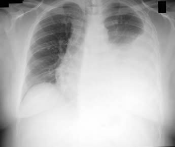
A pleural effusion is a collection of fluid in the space between the lung surface and the inside of the thorax. The tissue covering these surfaces is called the pleura, and a collection of fluid in an anatomical space is called an effusion, hence the term pleural effusion.
The main clinical conundrum where pleural effusions are concerned is working out the cause. There are lots of possible causes but most are rare. The most common differential diagnosis includes:
Less common causes include:
But let’s concentrate on the three most common. So how do you tell them apart? This includes a careful clinical history, an examination of the patient and eventually taking a sample of the fluid for laboratory analysis.
Clinical History
The first step in investigating any differential diagnosis is to take a full clinical history of the patient’s presenting complaint. It may sound too simple but more than 90% of diagnoses are made from the patient’s own story of what is happening; the examination and investigations are there to prove or disprove the most likely diagnosis.
All patients with pleural effusions eventually have shortness of breath, a cough and fast breathing (tachypnoea), but there are other factors in the history that may help decipher the underlying cause. For a pleural effusion the factors in the clinical history that might suggest a particular diagnosis are:
The main clinical conundrum where pleural effusions are concerned is working out the cause. There are lots of possible causes but most are rare. The most common differential diagnosis includes:
- Heart failure
- Infection (including TB)
- Malignancy
Less common causes include:
- Connective tissue disorders e.g. Lupus, rheumatoid disease
- Lymphatic abnormalities e.g. disruption of the thoracic duct allowing chyle to leak into the chest (chyle: Greek for “juice”, a milky bodily fluid consisting of lymph and emulsified fats. It is formed in the small intestine during digestion of fatty foods, and taken up by lacteals or lymph vessels)
- Iatrogenic causes (caused by doctors) e.g. damage to the oesophagus during endoscopy
- Movement of fluid from below the diaphragm e.g. severe ascites tracking up into the chest
But let’s concentrate on the three most common. So how do you tell them apart? This includes a careful clinical history, an examination of the patient and eventually taking a sample of the fluid for laboratory analysis.
Clinical History
The first step in investigating any differential diagnosis is to take a full clinical history of the patient’s presenting complaint. It may sound too simple but more than 90% of diagnoses are made from the patient’s own story of what is happening; the examination and investigations are there to prove or disprove the most likely diagnosis.
All patients with pleural effusions eventually have shortness of breath, a cough and fast breathing (tachypnoea), but there are other factors in the history that may help decipher the underlying cause. For a pleural effusion the factors in the clinical history that might suggest a particular diagnosis are:
Heart failure |
Known ischaemic heart disease, breathlessness on minimal exertion or when lying flat, coughing out frothy white sputum (oedema fluid from the lungs) |
Infection |
Sweats, chills, aches, chest pain, coughing out purulent sputum, chronic lung disease e.g. chronic obstructive pulmonary disease |
Malignancy |
Weight loss, coughing out blood (haemoptysis), smoker, exposure to asbestos or other carcinogens |
Clinical Examination
The clinical examination can help to add more weight to one diagnosis over another. The key points in the clinical examination include:
The clinical examination can help to add more weight to one diagnosis over another. The key points in the clinical examination include:
Heart failure |
Pitting oedema in the legs (fluid in the legs which when pushed leaves a temporary “dent” in the patients tissue), crackles at the lung bases not cleared by coughing (pulmonary oedema), raised jugular venous pressure |
Infection |
Fever, bronchial breathing (sounds from the bronchi and bronchioles being transmitted to the chest wall by consolidated lung) |
Malignancy |
Muscle wasting (cachexia) |
The problem with using clinical history and examination to differentiate the cause of a pleural effusion is that the diagnoses can overlap. Both heart failure and malignancy can predispose to infection, and because all three diagnoses are relatively common it is possible for a patient with heart failure to develop cancer even though it is unrelated to their ischaemic heart disease.
So the next step is to take some of the pleural fluid out of the patient and analyse it in a laboratory.
Laboratory analysis
Thoracocentesis is the technical term for the medical procedure where a needle is inserted into the pleural space and fluid withdrawn. If the effusion is so large that it is causing significant symptoms in the patient a drain may be left in place to remove more of the fluid, not just the small amount needed for further investigations.
At least three samples should be taken with about 20-25mls of fluid into three separate sterile universals. A quick look at the sample can spot if it is think and green and full of pus, in which case it is an empyema and the patient has an infection.
The samples are then sent to the biochemistry, microbiology and cytology laboratories.
Biochemistry
The first step in laboratory analysis of a pleural effusion is to decide if the fluid is a transudate or an exudate. A transudate occurs because the hydrostatic or osmotic fluid pressure in the blood stream or other body fluid increases forcing fluid into the lower pressure pleural space; transudates occur with heart failure. An exudate occurs due to inflammation or abnormal tissue drawing fluid into the pleural space; exudates occur with infection and malignancy.
Biochemistry laboratories do this by measuring the protein, cholesterol and Lactate Dehydrogenase (LDH) in the fluid. An exudate is defined as a pleural fluid that meets 1 of the 3 criteria:
If the values are close to the cut offs above then transudates and exudates may be misclassified so it is probably sensible to keep an open mind in these cases and maybe repeat the thoracocentesis if the diagnosis is unclear.
Microbiology
The microbiology laboratory will look at the number and type of white blood cells in the pleural fluid as well as doing a Gram’s stain for bacteria, a Ziehl-Neelson (ZN) stain for tuberculosis and a bacterial culture.
If the pleural fluid is full of neutrophil polymorphs and a bacterium is seen on microscopy then the patient has an infection; the diagnosis is pneumonia with or without an empyema. If the pleural fluid is full of lymphocytes and a mycobacterium is seen on ZN stain then the diagnosis is tuberculosis.
If no bacteria are seen the diagnosis is less certain and therefore a positive culture is required to confirm the presence of an infection, the bacteria most likely to cause an infected pleural effusion are Streptococcus pneumonia, Streptococcus anginosus, Staphylococcus aureus or Haemophilus influenzae. A positive mycobacterial culture clinches the diagnosis of tuberculosis but this usually takes up to two weeks.
But problems occur when the sample is full of lymphocytes and no organisms are seen… could this still be tuberculosis or is it cancer? If the diagnosis is urgent, i.e. treatment is needed as soon as possible other tests are required… bring on the very small speciality of cytology.
Cytology
The cytology laboratory looks at the types of cells in the pleural fluid sample and also looks to see if they have features of cancerous cells. The cytology laboratory can also help with the diagnosis of infection as they can see what type of white blood cells are present and whether they are reacting to a stimulus e.g. infection. They can also look for molecules on the cell walls which are associated with various diseases including cancer and connective tissue disorders.
Cytological analysis is not 100% for diagnosing cancer (in fact it’s between 60-75%) so there are still some patients where the differential diagnosis is still tuberculosis or cancer.
Other tests
So this brings me back to the question from the respiratory doctors about what is the chemical test that can help diagnose tuberculosis?
It turns out that there is another biochemical test which can help distinguish between tuberculosis and malignancy but only in the context of a lymphocytic exudative pleural effusion. The test is for adenosine deaminase (ADA). I don’t think this is a particularly new test but I’d never heard of it.
ADA is an enzyme involved in the breakdown of the nucleotide adenine (via adenosine). ADA is produced when lymphocytes are activated in the immune response to intracellular infections. Mycobacterium tuberculosis is an intracellular bacterium and so in tuberculosis ADA is produced in large amounts; whereas in malignancy it isn’t.
ADA in pleural fluid can be measured and is much quicker than culture for tuberculosis or cytology for malignancy. A level >40U/L is suggestive of tuberculosis. It is important to consider the result in the context of the patient as false positives and negatives can occur (no test in medicine is 100% reliable) but it can help support the clinical suspicion of one disease over another.
So the patient underwent a thoracocentesis and as expected the biochemistry results showed an exudate. Microbiology confirmed the presence of lymphocytes but the Gram stain and ZN stain showed no bacteria. The cytology also showed lymphocytes. Whilst waiting for the tuberculosis culture and further cytology results the biochemists did the ADA test which showed a high ADA and the respiratory doctors made a diagnosis of tuberculosis and started treatment. Two weeks later the Microbiologists grew a mycobacterium which eventually confirmed the diagnosis of tuberculosis but we were much slower than the biochemists! Plenty of time for the Microbiologist to have an extra cup of tea and a biscuit!!! "If only there was a staff room…" I hear you cry!
So the next step is to take some of the pleural fluid out of the patient and analyse it in a laboratory.
Laboratory analysis
Thoracocentesis is the technical term for the medical procedure where a needle is inserted into the pleural space and fluid withdrawn. If the effusion is so large that it is causing significant symptoms in the patient a drain may be left in place to remove more of the fluid, not just the small amount needed for further investigations.
At least three samples should be taken with about 20-25mls of fluid into three separate sterile universals. A quick look at the sample can spot if it is think and green and full of pus, in which case it is an empyema and the patient has an infection.
The samples are then sent to the biochemistry, microbiology and cytology laboratories.
Biochemistry
The first step in laboratory analysis of a pleural effusion is to decide if the fluid is a transudate or an exudate. A transudate occurs because the hydrostatic or osmotic fluid pressure in the blood stream or other body fluid increases forcing fluid into the lower pressure pleural space; transudates occur with heart failure. An exudate occurs due to inflammation or abnormal tissue drawing fluid into the pleural space; exudates occur with infection and malignancy.
Biochemistry laboratories do this by measuring the protein, cholesterol and Lactate Dehydrogenase (LDH) in the fluid. An exudate is defined as a pleural fluid that meets 1 of the 3 criteria:
- Protein >2.9g/L
- Cholesterol >45mg/L (1.165 mmol/L)
- LDH >1.45x the laboratory upper limit for serum LDH
If the values are close to the cut offs above then transudates and exudates may be misclassified so it is probably sensible to keep an open mind in these cases and maybe repeat the thoracocentesis if the diagnosis is unclear.
Microbiology
The microbiology laboratory will look at the number and type of white blood cells in the pleural fluid as well as doing a Gram’s stain for bacteria, a Ziehl-Neelson (ZN) stain for tuberculosis and a bacterial culture.
If the pleural fluid is full of neutrophil polymorphs and a bacterium is seen on microscopy then the patient has an infection; the diagnosis is pneumonia with or without an empyema. If the pleural fluid is full of lymphocytes and a mycobacterium is seen on ZN stain then the diagnosis is tuberculosis.
If no bacteria are seen the diagnosis is less certain and therefore a positive culture is required to confirm the presence of an infection, the bacteria most likely to cause an infected pleural effusion are Streptococcus pneumonia, Streptococcus anginosus, Staphylococcus aureus or Haemophilus influenzae. A positive mycobacterial culture clinches the diagnosis of tuberculosis but this usually takes up to two weeks.
But problems occur when the sample is full of lymphocytes and no organisms are seen… could this still be tuberculosis or is it cancer? If the diagnosis is urgent, i.e. treatment is needed as soon as possible other tests are required… bring on the very small speciality of cytology.
Cytology
The cytology laboratory looks at the types of cells in the pleural fluid sample and also looks to see if they have features of cancerous cells. The cytology laboratory can also help with the diagnosis of infection as they can see what type of white blood cells are present and whether they are reacting to a stimulus e.g. infection. They can also look for molecules on the cell walls which are associated with various diseases including cancer and connective tissue disorders.
Cytological analysis is not 100% for diagnosing cancer (in fact it’s between 60-75%) so there are still some patients where the differential diagnosis is still tuberculosis or cancer.
Other tests
So this brings me back to the question from the respiratory doctors about what is the chemical test that can help diagnose tuberculosis?
It turns out that there is another biochemical test which can help distinguish between tuberculosis and malignancy but only in the context of a lymphocytic exudative pleural effusion. The test is for adenosine deaminase (ADA). I don’t think this is a particularly new test but I’d never heard of it.
ADA is an enzyme involved in the breakdown of the nucleotide adenine (via adenosine). ADA is produced when lymphocytes are activated in the immune response to intracellular infections. Mycobacterium tuberculosis is an intracellular bacterium and so in tuberculosis ADA is produced in large amounts; whereas in malignancy it isn’t.
ADA in pleural fluid can be measured and is much quicker than culture for tuberculosis or cytology for malignancy. A level >40U/L is suggestive of tuberculosis. It is important to consider the result in the context of the patient as false positives and negatives can occur (no test in medicine is 100% reliable) but it can help support the clinical suspicion of one disease over another.
So the patient underwent a thoracocentesis and as expected the biochemistry results showed an exudate. Microbiology confirmed the presence of lymphocytes but the Gram stain and ZN stain showed no bacteria. The cytology also showed lymphocytes. Whilst waiting for the tuberculosis culture and further cytology results the biochemists did the ADA test which showed a high ADA and the respiratory doctors made a diagnosis of tuberculosis and started treatment. Two weeks later the Microbiologists grew a mycobacterium which eventually confirmed the diagnosis of tuberculosis but we were much slower than the biochemists! Plenty of time for the Microbiologist to have an extra cup of tea and a biscuit!!! "If only there was a staff room…" I hear you cry!
 RSS Feed
