The patient was a 42 year old man who presented to his GP with a fever and severe pain in his axilla. On examination there was tender lymphadenopathy with overlying erythema; the largest lymph node measured 4cm. Further questioning elicited that the patient had a crop of painful, erythematous vesicles distally on the same arm. The GP wondered if this might be a localised skin infection with something like Staphylococcus aureus, the most common cause of skin infections; the lymph node involvement might represent regional lymphatic spread e.g. lymphangitis. The GP prescribed Flucloxacillin and suggested that the patient should return if there was no improvement in a few days.
Three days later the patient was no better and returned to his GP. The GP re-examined the patient and noted some minor scratches on the patient’s hands and arms. The patient explained that he had adopted a female kitten who liked to play with his hands, batting him without always remembering to retract her claws. As a result the patient had sustained a few nasty scratches when the play became a little “rough”.
Three days later the patient was no better and returned to his GP. The GP re-examined the patient and noted some minor scratches on the patient’s hands and arms. The patient explained that he had adopted a female kitten who liked to play with his hands, batting him without always remembering to retract her claws. As a result the patient had sustained a few nasty scratches when the play became a little “rough”.
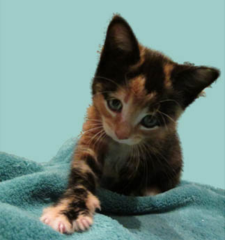
"Granola"
The GP rang the Microbiologist at the local hospital to discuss whether this might be an infection acquired from the kitten, the Microbiologist said it sounded like “cat scratch disease” … "oh honestly" said the GP knowing the reputation for joking around by the Microbiologist. "No, no honest" said the Microbiologist, “it really is likely to be cat scratch disease”. “oh there really is something called cat scratch disease then, I thought you were joking!?” said the GP.
What is cat scratch disease (CSD)?
CSD is an infection caused by the bacterium Bartonella henselae. This bacterium does not cause disease in the cat but it carries it inside its red blood cells for years; 90% of cats under 1 year have evidence of past B. henselae infection with about 50% having ongoing bacteraemia. Infection in humans occurs when the bacterium is inoculated through the skin usually as a result of a scratch from an infected cat’s claw… hence the name? Infection can also occur through the bite of cat fleas infected with B. henselae; this is the main route of transmission from cat-to-cat (I’d need to ask a vet if it is better known as Flea-Bite Fever in cats, although it doesn’t sound as good as Cat Scratch Disease).
Infection occurs approximately 3-10 days after the initial scratch at the site of inoculation. A localised skin reaction develops with vesicles, erythema then papules. This localised reaction usually lasts for up to 3 weeks and then resolves completely with no scarring. Very occasionally the initial skin reaction can persist for a few months.
The classic feature of CSD is regional lymphadenopathy that occurs proximal to the original inoculation site about 2 weeks after the initial scratch. The lymph nodes are usually up to 5cm in diameter but have been known to get as large as 10cm. They are tender and there is often overlying erythema. Occasionally the lymph nodes suppurate and discharge to the skin like large abscesses. Lymphadenopathy usually settles spontaneously within 4 months but can occasionally persist for longer.
In 5-10% of patients B. henselae disseminates to involve the liver, spleen, eye, central nervous system, muscles, joints or the heart.
Splenomegaly is common (10-12%); liver granulomas can also occur but less commonly. The eyes can be affected either by localised inoculation causing conjunctivitis, conjunctival granulomas and preauricular lymphadenopathy, or by neuroretinitis and associated loss, or partial loss, of vision. Encephalopathy can occur up to 6 weeks after infection which can involve confusion or coma, seizures and focal neurological signs; this usually resolves with a few weeks but some patients will have residual neurological defects. Muscles and joint pains can occur to the point of being severe and disabling until they spontaneously resolve. B. henselae is a cause of culture-negative infective endocarditis but this is very rare.
How to diagnose CSD
CSD is suspected with an appropriate history of contact with cats and the classic findings of localised skin reaction and regional lymphadenopathy. The most common method to try and prove the diagnosis is serology because B. henselae is fastidious and doesn’t grow with conventional techniques in the microbiology laboratory. Serology is prone to false positives and because it detects IgG which persists after infection resolves, it may indicate past rather than acute infection. Having said that rising Bartonella IgG titres, or a value >256, in the context of the classical skin and lymph node signs is good enough to make the diagnosis. If the classical skin and lymph node signs are absent the diagnosis requires further investigation using different techniques.
PCR is now the method of choice for diagnosing disseminated B. henselae infection. Almost any body tissue or fluid can be tested however the sensitivity is only 45-75% so it is still far from perfect. Histology can also suggest a possible diagnosis of CSD but again is not 100%. Because of the limitations with the diagnostic tools available, CSD is often suspected but never actually proven. If there is no other plausible explanation for the patient’s condition then it is probably worth managing as CSD in these situations.
How is CSD treated?
There is little evidence that antibiotics make much difference in the classical skin and lymphadenopathy form of CSD and they should therefore not be given. Patients should be reassured that this infection will settle on its own. There are some experts who do give macrolides like Clarithromycin or Azithromycin to these patients based upon a single study of 29 patients who were randomised to either receive Azithromycin or not. The treatment group had faster reduction in size of lymphadenopathy but there was no difference in overall response. A non-randomised study of 268 patients treated with various antibiotics or no antibiotics showed little difference in rates of dissemination or final outcome. Unfortunately there is no way to predict which infections will disseminate.
Disseminated infection is a different matter and all experts agree that disseminated B. henselae infection SHOULD be treated with antibiotics. Azithromycin PLUS Rifampicin for 10-14 days is commonly used for most disseminated disease with Doxycycline PLUS Rifampicin being used to treat neurological disease and neuroretinitis in adults. Neuroretinitis requires 4 to 6 weeks of treatment whichever regimen is being used.
So what happened to the patient? Well nothing really… he woke up and realised that he was a Consultant Microbiologist who had just had a bad dream as a result of an over active imagination and having taken on a new member of the Microbiology Nuts & Bolts team… a 7 week old tortoiseshell kitten called “Granola”. Granola has taken up a massive amount of our time and that’s why we never quite got around to writing a blog last week… that and the number of scratches on our hands and feet from an overly playful kitten!
P.S. All of the information about CSD is correct though :-)
 RSS Feed
