The Microbiologist groaned as his pager went off at midnight, waking him after he had just dozed off. He stumbled over the sleeping cat and went to answer the call, while his wife stirred and uttered something unrepeatable. However, rather than one of the normal “you should know this from medical school” types of call the Microbiologist soon snapped awake as the story unfolded.
The patient had returned 5 days ago from visiting family in Israel. They had a fever and muscle pains but had come in to hospital as they had noticed a florid rash all over their body when getting undressed to go to bed. The A&E doctor was concerned this might be meningococcal sepsis but as the patient was an adult they thought they better get advice about what to do.
The patient had returned 5 days ago from visiting family in Israel. They had a fever and muscle pains but had come in to hospital as they had noticed a florid rash all over their body when getting undressed to go to bed. The A&E doctor was concerned this might be meningococcal sepsis but as the patient was an adult they thought they better get advice about what to do.

The Microbiologist agreed that meningococcal sepsis should be part of the differential diagnosis but also asked the doctor to go and ask and look for any funny lumps or bumps on the patients skin, especially any black lumps about half-a-centimetre in size. A few minutes later the doctor came back to the phone saying there were a few such black lesions on the back of the patient’s legs.
“This could be the rickettsioses Mediterranean Spotted Fever” suggested the Microbiologist, “but you ought also to treat for meningococcal sepsis until we know for sure”.
The patient was started on Doxycycline for Mediterranean Spotted Fever and Ceftriaxone for the possible alternative diagnosis of meningococcal sepsis.
As the Microbiologist climbed back in to bed his wife asked with a groan, “sensible call?”
“Yep, very sensible” replied the Microbiologist, but his wife was already asleep again and didn’t hear him.
So why do Microbiologists sometimes think of zebras when they hear hooves rather than horses… well let’s put it another way… if you’re on a safari in Africa and you hear hooves what’s it most likely to be, a horse or a zebra? Rare infections become more common if the context in which you are seeing a patient changes, for example malaria in someone who has never left the UK is very unlikely whereas malaria in someone who has just returned from a safari would be much more likely.
What are the rickettsioses?
The rickettsioses are a group of similar bacterial infections, usually transmitted by tick bites. There are many different types of rickettsioses but for this blog I am going to concentrate on the three most “commonly” seen in the UK, although, in the UK, any type of rickettsial infection is an uncommon diagnosis. In order of severity these are: Rocky Mountain Spotted Fever (RMSF caused by Rickettsia rickettsi), Mediterranean Spotted Fever (MSF caused by Rickettsia conorii) and African Tick Bite Fever (ATBF caused by Rickettsia africae).
How do rickettsioses present?
Most rickettsial infections present with a fever, rash and a vasculitis however the clinical features and severity depends on which bacterium the patient is infected with. The vasculitis can cause multi-organ damage including encephalitis, pulmonary oedema, adult respiratory distress syndrome, cardiac arrhythmias, coagulopathies, gastrointestinal bleeding, liver and renal failure and skin necrosis. The incubation period from tick bite to symptoms ranges from 2 -21 days.
RMSF can be mild if treated early or severe and fatal, if untreated. The original tick bite is usually not apparent. It presents with fever, malaise, muscle pain, headache and conjunctival redness. A maculopapular rash appears on the hands and feet of only about 30% of patients 3-4 days into the illness, and then spreads to the rest of the body; yep that leaves 70% of patients with Rocky Mountain Spotted Fever who do not have the spots! The rash becomes petechial around day 6 as the vasculitis develops which then causes end organ damage. Mortality ranges from 20-80% without treatment, however with treatment, started within 5 days of illness, almost all patients will survive.
MSF can be mild or severe. There is often an eschar at the site of the tick bite; a raised red lesion up to 5mm in size with a black necrotic centre. A maculopapular rash usually appears all over the body on day 4-5 in 97-99% of patients. The untreated mortality is 10-30%. Severe infections are associated with cardiac, neurological and renal damage.
ATBF is similar to MSF but is usually only a mild infection. An eschar is usually present and the fever, myalgia and headache are abrupt in onset. Only about 50% of patients have a maculopapular rash. Complications are rare and symptoms usually settle after 10 days even without treatment. Antibiotics can hasten the resolution of symptoms. Deaths are very rare.
Where do you catch rickettsia?
RMSF occurs throughout North, Central and much of South America. Most cases occur in the USA, Canada, Mexico, Columbia, Brazil and Argentina. It was called Rocky Mountain Spotted Fever because it was first described in the area of the Rocky Mountains in the USA. RMSF is transmitted by American dog ticks, Rocky Mountain wood ticks, brown dog ticks and cayenne ticks. (I assume the tick is brown, not necessarily the dog!?)
MSF occurs in areas of Europe, Africa and the Middle East surrounding the Mediterranean, Caspian and Black Seas. MSF is transmitted by the brown dog tick, which we do have in the UK but it doesn't have the rickettsial bacteria.
ATBF occurs in Sub-Saharan Africa including South Africa, Botswana, Swaziland and Zimbabwe. ATBF is transmitted by the African bont tick and the tropical bont tick. (I have no idea what a bont is, if you know then please let me know as well)
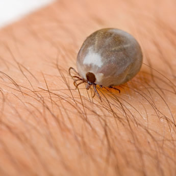
Brown dog tick
There is no transmission of rickettsia from person-to-person although “outbreaks” can occur in contacts if all the cases have a common exposure to tick bites e.g. family holiday.
How are rickettsioses diagnosed?
The diagnosis of rickettsioses is initially based on clinical suspicion. Do not wait for blood tests before starting treatment.
The most common method for confirming a rickettsial infection is by detecting a 4 fold rise in IgG antibody between serum samples taken acutely (on presentation) and in convalescence (2-3 weeks after presentation). This type of investigation is detecting the body’s ability to produce specific antibody against an infection after exposure; the amount of antibody increases as the body becomes better at producing it. It takes 2-3 weeks for the body to produce a 4 fold rise in titre and RMSF tends to kill patients in 8 days, this is why a clinical diagnosis has to be made and treatment started before the diagnosis is confirmed. Remember, a negative result in the acute serum does not rule out rickettsia infection, a repeat test at 2-3 weeks is needed.
In an acute infection it is sometimes possible to detect rickettsia bacteria in blood by PCR although again a negative PCR does not rule out the infection.
How are rickettsioses treated?
The treatment of rickettsial infection is Doxycycline, either IV or oral depending on severity of infection. Even in pregnancy and children the treatment is Doxycycline. Doxycycline is by far the best treatment of rickettsial infections and the theoretical risk of dental staining and bone developmental abnormalities have not been seen in clinical use (see previous blog).
Adults: IV or PO Doxycycline 100mg BD
Children: IV or PO Doxycycline 2.2mg/kg BD
If there is a definite history of severe Doxycycline allergy then Chloramphenicol is the alternative however it is not as effective and it is associated with a higher mortality.
Duration: until 3 days after fever settles, usually 5-10 days total course.
So the patient was started on antibiotics and serum and blood were sent off to the Reference Laboratory for rickettsioses. Blood cultures and an EDTA blood for PCR were taken to look for meningococcal sepsis. By the following day the patient was starting to feel a bit better. Three days after admission the Microbiologist got a call from the Reference Laboratory saying that they had detected Rickettsia in the blood sample. The blood cultures and meningococcal PCR were negative. The patient continued on Doxycycline but the Ceftriaxone was stopped as the patient did not have meningococcal sepsis. Five days after starting treatment the patients fever settled and the Doxycycline was continued for a further three days (8 days of treatment). At follow up 3 weeks later a repeat blood test showed a greater than 4 fold rise in IgG to rickettsia and the diagnosis was confirmed. Even the Microbiologist’s wife was slightly impressed at the diagnosis…!
 RSS Feed
