The Microbiologist gave a big sigh. It was a beautiful spring day outside, perfect for hitting the forest trails, and here he was sat at the duty desk advising on how to treat athletes foot… he sighed again…
The phone rang and with a heavy heart the Microbiologist answered.
“I have a patient who I think has a fungal infection” said the voice on the other end of the line.
“Here we go again” thought the Microbiologist.
“What kind of fungal infection?”
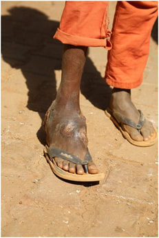
“I’m not really sure. He has a lump on his foot which has been getting worse since he came back from Africa a few months ago” said the voice.
The Microbiologist sat up straight. This was more like it, something weird and wonderful.
“Who am I speaking to? What was your patient doing in Africa? Did he hurt himself, cuts to his feet, anything like that?”
“Oh sorry, it’s the Dermatologist here, busy clinic and forgot my manners. The patient was volunteering, helping to build a school in a rural village, he was there for a long time and I suspect as he was wearing flip-flops a lot, he had many scratches and cuts to his feet during that time. I thought it might be another case of sporotrichosis, but there is no sporotrichoid spread and the lump is completely painless. There does look to be a sinus forming as well. What do you think?”
“It does sound like eumycetoma. Here’s what I think we should do…”
The phone rang and with a heavy heart the Microbiologist answered.
“I have a patient who I think has a fungal infection” said the voice on the other end of the line.
“Here we go again” thought the Microbiologist.
“What kind of fungal infection?”
“I’m not really sure. He has a lump on his foot which has been getting worse since he came back from Africa a few months ago” said the voice.
The Microbiologist sat up straight. This was more like it, something weird and wonderful.
“Who am I speaking to? What was your patient doing in Africa? Did he hurt himself, cuts to his feet, anything like that?”
“Oh sorry, it’s the Dermatologist here, busy clinic and forgot my manners. The patient was volunteering, helping to build a school in a rural village, he was there for a long time and I suspect as he was wearing flip-flops a lot, he had many scratches and cuts to his feet during that time. I thought it might be another case of sporotrichosis, but there is no sporotrichoid spread and the lump is completely painless. There does look to be a sinus forming as well. What do you think?”
“It does sound like eumycetoma. Here’s what I think we should do…”
What the heck is eumycetoma?
Eumycetoma are fungal infections which initially start in the skin but can erode into deeper tissue. They most commonly occur in tropical countries including Sudan, Senegal, Mauritania, Niger, Kenya, Mali, Cameroon, Somalia, Djibouti, India, Iran, Venezuela, Argentina, Colombia and Chile.
The fungi that cause eumycetoma are environmental moulds usually found in soil and decaying plant matter. The most common, causing >90% of eumycetoma, are:
How does eumycetoma occur?
The first step in the development of eumycetoma is the inoculation of fungal spores into tissue, usually from a puncture wound with a thorn. This explains why eumycetomas are more common on the feet and legs as these are the most common sites for inoculation (approximately 90%). They are also more common in agricultural workers and outdoor labourers as well as where shoes are not commonly worn. There is no person-to-person transmission.
However, if inoculation were the only predisposing factor, you’d expect eumycetoma would be very common; yet weirdly it is actually relatively uncommon. It is suspected that there is a genetic predisposition to developing eumycetoma, including problems with neutrophil function and cell mediated immunity, meaning the patient is unable to clear the fungus once it has been inoculated.
How does eumycetoma present?
Eumycetoma initially presents with a small painless nodule or nodules, which slowly enlarge and become tethered to the underlying tissues. As the nodule enlarges a sinus tract develops and the lesion starts to discharge pus containing granules which contains fungal mycelia. Slowly the lesion gets bigger and invades deeper causing tissue destruction and significant deformity. This is especially true where healthcare systems are remote, not available or unaffordable. It can be difficult to distinguish eumycetoma from sarcomas (a type of soft tissue cancer) without further investigation.
Eumycetoma are fungal infections which initially start in the skin but can erode into deeper tissue. They most commonly occur in tropical countries including Sudan, Senegal, Mauritania, Niger, Kenya, Mali, Cameroon, Somalia, Djibouti, India, Iran, Venezuela, Argentina, Colombia and Chile.
The fungi that cause eumycetoma are environmental moulds usually found in soil and decaying plant matter. The most common, causing >90% of eumycetoma, are:
- Madurella mycetomatis
- Nigrograna mackinnonii
- Trematosphaeria grisea
- Scedosporum apiospermum (Pseudalelescheria boydii)
- Falciformispora senegalensis
How does eumycetoma occur?
The first step in the development of eumycetoma is the inoculation of fungal spores into tissue, usually from a puncture wound with a thorn. This explains why eumycetomas are more common on the feet and legs as these are the most common sites for inoculation (approximately 90%). They are also more common in agricultural workers and outdoor labourers as well as where shoes are not commonly worn. There is no person-to-person transmission.
However, if inoculation were the only predisposing factor, you’d expect eumycetoma would be very common; yet weirdly it is actually relatively uncommon. It is suspected that there is a genetic predisposition to developing eumycetoma, including problems with neutrophil function and cell mediated immunity, meaning the patient is unable to clear the fungus once it has been inoculated.
How does eumycetoma present?
Eumycetoma initially presents with a small painless nodule or nodules, which slowly enlarge and become tethered to the underlying tissues. As the nodule enlarges a sinus tract develops and the lesion starts to discharge pus containing granules which contains fungal mycelia. Slowly the lesion gets bigger and invades deeper causing tissue destruction and significant deformity. This is especially true where healthcare systems are remote, not available or unaffordable. It can be difficult to distinguish eumycetoma from sarcomas (a type of soft tissue cancer) without further investigation.
- Very occasionally eumycetoma can spread through the lymphatics or blood stream to cause disseminated infection.
How do you diagnose a eumycetoma?
A clinical diagnosis of eumycetoma can be made based on the presence of a painless lump, a sinus tract and discharging black granules (other colour granules can be bacterial, especially actinomycosis).
In order to identify the causal fungus, tissue, aspirate or grains should be cultured on Sabouraud dextrose agar at both room temperature and 37oC. Culture can take 6-8 weeks to be positive.
Important Note: When plating out tissue don’t grind it to make it easier to spread as this can damage the fungus and stop it growing. A faster method is to use PCR to identify the fungus but this is a Reference Laboratory test and not widely available.
Histopathological examination of tissue, aspirate or grains can also identify fungal mycelia and a granulomatous reaction, although an experienced eye is needed to distinguish the different species of fungi.
How is eumycetoma treated?
There have been no clinical trials looking at the treatment of eumycetoma, although current recommendations are below and require identification of the causative fungus first.
Causative Fungus |
Treatment |
|
PO Itraconazole 200mg BD |
|
PO Voriconazole 400mg BD for 3 doses THEN 200mg BD |
Note: the common antifungals Amphotericin B and echinocandins like Caspofungin have no activity against the fungi causing eumycetoma and should not be used.
Surgery can be considered for early small nodules but for larger lesions there is little additional benefit over antifungals alone.
There is no specific duration of treatment. Courses over 12 months long are often required. The basic rule of thumb is that treatment should be continued until at least 3 months after clinical and radiological “cure”.
So the Dermatologist went back to the patient and aspirated some fluid from the patient’s lesion which was shown to contain fungal hyphae. A sample was sent for PCR which confirmed the presence of Madurella mycetomatis. Because the eumycetoma was small it was decided to surgically remove it and the patient was started on Itraconazole.
It was a good lesson for the Microbiologist. Not all fungal feet are dull, some are weird and exciting… honest!
P.S. Wear proper shoes ECIC! [Nah, Humbug – free your feet!!]
 RSS Feed
